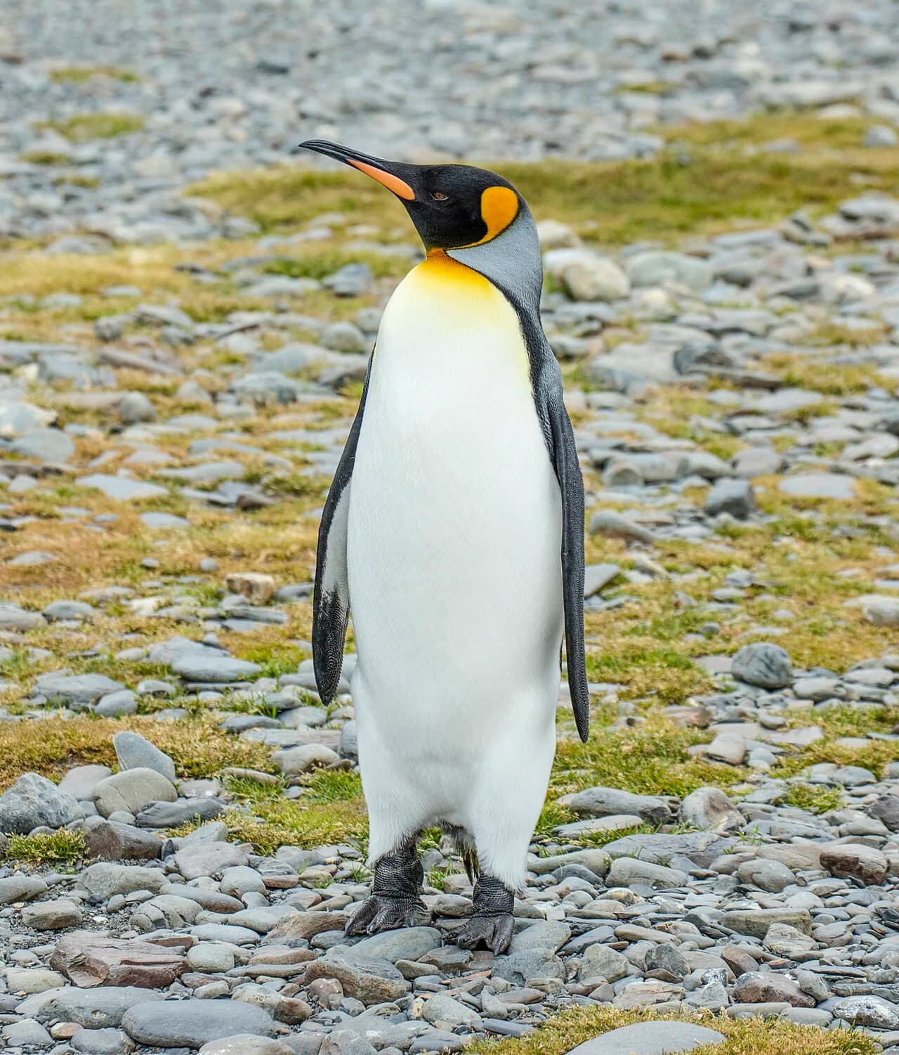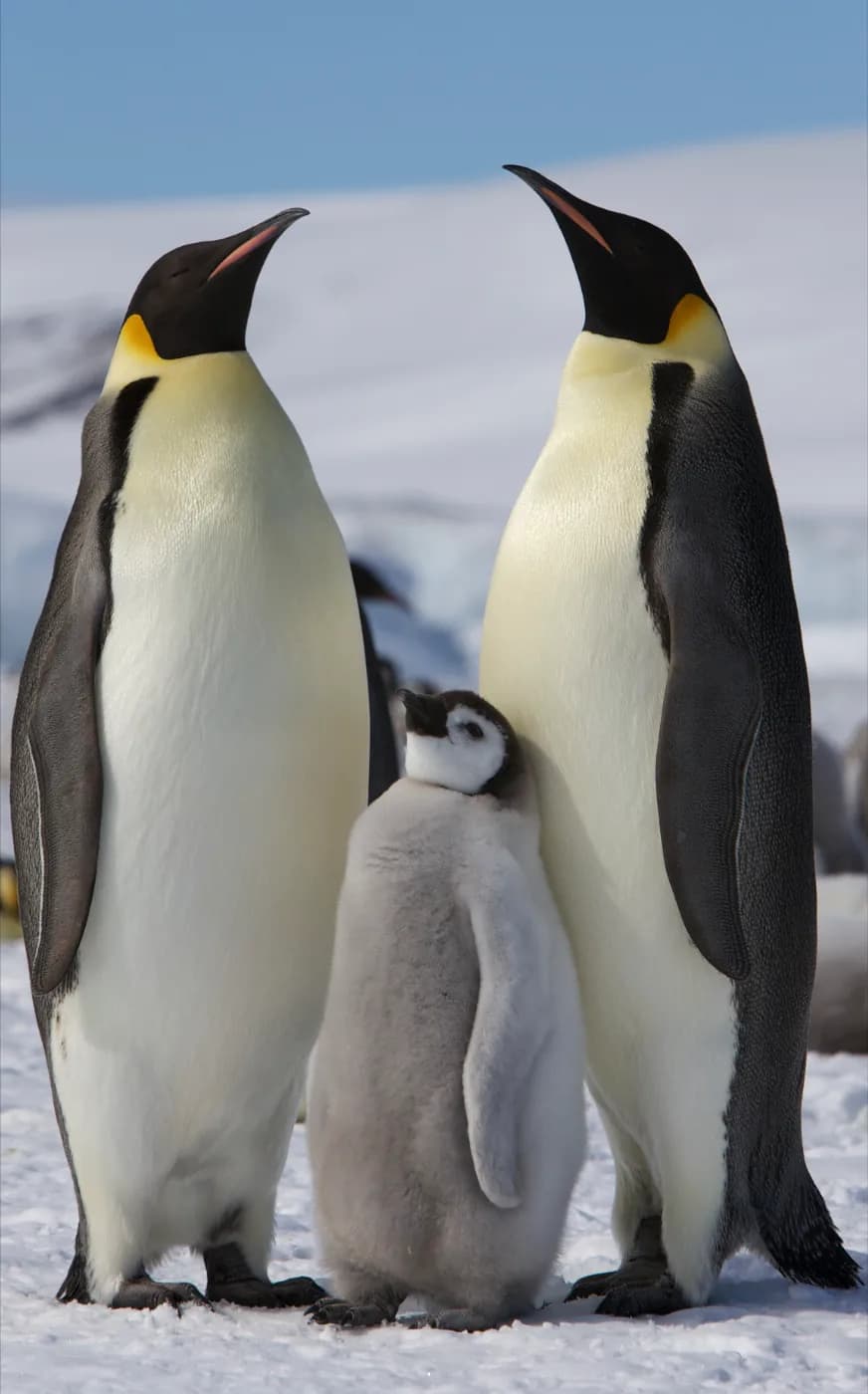
Look at a standing penguin and its feet seem to emerge straight from the bottom of its body. The thigh and knee are hidden farther up, beneath the feathers. Each flipper conceals another familiar structure: the framework of a bird's wing.
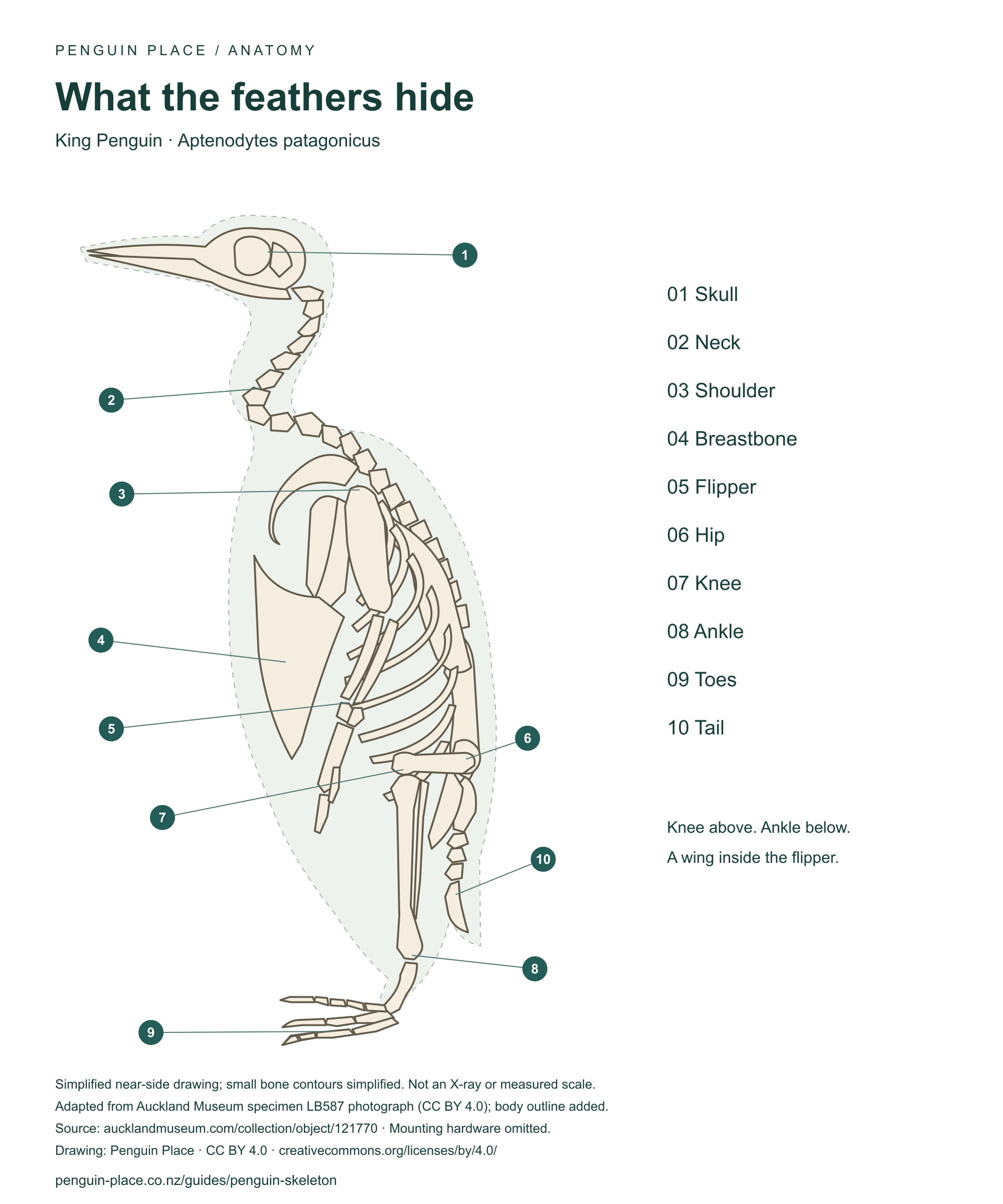
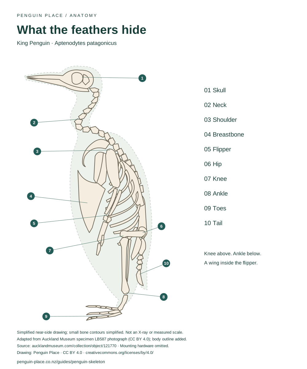
The illustration follows a King Penguin skeleton, specimen LB587, in Auckland Museum. Follow the bones from the hip to the toes, then from the shoulder to the flipper tip. The drawing makes those hidden limbs easier to recognise.1
The knees are there. The feathers hide them.
Find the hip towards the back of the skeleton. Now follow the short thigh bone forwards. Where it meets the lower leg is the knee: landmark 7 on the diagram. It sits well above the ankle, inside the outline we recognise as the penguin's body.
The joint down near the foot is easy to mistake for a knee bending the wrong way. That lower joint is the ankle. The knee sits higher, hidden by soft tissues and feathers. Penguins have the same basic leg sequence as other birds, with proportions and a posture that look unfamiliar from the outside.2
- Hip → femur (thigh)
- Knee → tibiotarsus, with the fibula alongside
- Ankle → tarsometatarsus (fused foot region)
- Toe bones → the ground
Working downwards from the hip, the femur forms the thigh. Below the knee, the main lower-leg bone is the tibiotarsus, with the narrower fibula alongside it. Below the ankle sits the tarsometatarsus, a short foot element formed from fused ankle and foot bones, leading to the toes.3
For a shorter version of this answer, see do penguins have knees?.
And the waddle?
A penguin's walk can look inefficient, but appearance is a poor guide to the mechanics. In experiments with emperor penguins, researchers found that the side-to-side movement helped conserve mechanical energy. Their explanation for the high cost of walking instead emphasised how rapidly short legs must generate muscular force.4
Walking still costs emperor penguins considerable energy. The rocking motion helps recover mechanical energy between steps; the study does not establish that every penguin species walks in the same way.4
Inside the flipper is a wing
Follow the flipper from the shoulder. First comes a single upper-wing bone, the humerus. Beyond the elbow are two forearm bones, the radius and ulna. Farther out sit the wrist and hand region, followed by finger bones.
A flipper is a modified bird wing. Its bones and joints have been reshaped into a flattened swimming limb.5
- Shoulder → humerus (upper wing)
- Elbow → radius and ulna (forearm)
- Wrist → carpometacarpus (fused hand region)
- Finger bones → the flipper tip
Some bones within the hand region are fused, while other connections remain joints. Anatomical studies describe restricted motion at penguin wing joints; observations of swimming birds show that the flipper can still bend and twist.5
For the broader flight question, see why penguins cannot fly through the air. Their ability to work underwater is explored further in how penguins dive so deep.
The chest gives the muscles somewhere to pull
Bones do not move themselves. Muscles need attachment points, and the chest and shoulder girdle provide them.
On the drawing, the broad breastbone lies in front of the ribs. The shoulder region includes the scapula, or shoulder blade, and supporting bones that connect the wing to the trunk. A penguin's scapula is expanded compared with that of many flying birds, providing an attachment area for a muscle involved in lifting the wing.6
That lifting movement matters underwater. A flipper can produce thrust during both halves of its stroke, so the return movement is part of propulsion rather than simply resetting the wing. Water places very different demands on the limb from air.6
Denser bones, not a body made of solid rods
Penguin bones are often described simply as solid. Microscopic sections of their limb bones show thick walls of compact bone and reduced internal cavities, with variation between bones and across penguin lineages. Dense bone still has an internal structure; it is not a uniform solid rod.7
These cross-sections describe bone structure. How long a bird can stay underwater also depends on how it stores and uses oxygen, explored in how penguins dive so deep.
Put the feathers back
Switch the main diagram back to Body + bones. The knee sits inside the shaded outline, and the curved neck bones fit within a fuller shape of soft tissues and feathers. Muscles, plumage and posture all contribute to the outline of a living bird. The silhouette here is schematic, rather than a measurement of specimen LB587 in life.
To explore the outer coat, follow the feather and insulation guide, or look at how penguins grip food without teeth.
Bone names, without the jargon
This key pairs familiar body parts with their anatomical names. It covers the main structures shown in the drawing. A complete bone count would need to specify the specimen and how fused elements were counted.
- Skull and lower jawCranium and mandible
- Head; the outer beak covering is not bone
- Neck bonesCervical vertebrae
- The curve between head and chest
- Shoulder bladeScapula
- Shoulder girdle, beside the upper ribs
- BreastboneSternum
- Broad plate in front of the rib cage
- Upper-wing boneHumerus
- Shoulder to elbow
- Forearm bonesRadius and ulna
- Two bones beyond the elbow
- Fused hand regionCarpometacarpus
- Towards the flipper tip
- Thigh boneFemur
- Hip to knee
- Main lower-leg boneTibiotarsus
- Knee to ankle, with the fibula alongside
- Fused foot regionTarsometatarsus
- Ankle to toes
- Toe bonesPedal phalanges
- The digits at the foot
The terminology follows anatomical descriptions and the researcher-labelled King Penguin reference.235
Take the diagram with you
The downloads include the species, numbered key, source credit and a link back to this guide. All three are made from the same illustration shown above.
- Download the high-resolution labelled diagram (PNG)
- Download the printable diagram (PDF)
- Download the editable illustration (SVG)
The adapted illustration is available under CC BY 4.0. Credit Auckland Museum for the specimen photograph and Penguin Place for the drawing and annotations; indicate any further changes.
How this illustration was made
The drawing simplifies the near-side structures visible in Auckland Museum specimen LB587. Mounting wires, the supporting rod and the display base were omitted. Far-side limbs are left out to reduce overlap, and small bone contours are simplified. The body outline is a schematic addition; the positions are not a movement study, and the drawing is not to measured scale.
Names and relationships were cross-checked against published anatomical descriptions. You can inspect the museum's original specimen photograph and catalogue record alongside it.1
References
-
Auckland Museum Tāmaki Paenga Hira. Aptenodytes patagonicus, specimen LB587. Photograph licensed CC BY 4.0. https://www.aucklandmuseum.com/collection/object/121770 ↩ ↩2
-
Ksepka, D.T. (2011). A Map for Our Tour of the Penguin Skeleton. Researcher-labelled King Penguin skeleton. https://fossilpenguins.wordpress.com/2011/10/04/a-map-for-our-tour-of-the-penguin-skeleton/ ↩ ↩2
-
Ksepka, D.T. (2011). Tour of the Penguin Skeleton II: The Tarsometatarsus. https://fossilpenguins.wordpress.com/2011/06/06/tour-of-the-penguin-skeleton-ii-the-tarsometatarsus/ ↩ ↩2
-
Griffin, T.M. & Kram, R. (2000). Penguin waddling is not wasteful. Nature 408: 929. https://doi.org/10.1038/35050167 ↩ ↩2
-
Krahl, A. et al. (2023). Deep-time invention and hydrodynamic convergences through amniote flipper evolution. The Anatomical Record. https://doi.org/10.1002/ar.25119 ↩ ↩2 ↩3
-
Ksepka, D.T. (2010). Tour of the Penguin Skeleton I: Scapula. https://fossilpenguins.wordpress.com/2010/09/23/tour-of-the-penguin-skeleton-i-scapula/ ↩ ↩2
-
Ksepka, D.T., Werning, S., Sclafani, M. & Boles, Z.M. (2015). Bone histology in extant and fossil penguins (Aves: Sphenisciformes). Journal of Anatomy 227: 611–630. https://doi.org/10.1111/joa.12367 ↩